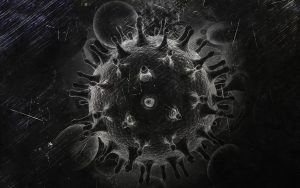
In the human medicine field, one of the earliest works utilized NIR spectra for detection of HIV-1 virus in plasma (Sakudo et al. 2005). The results yielded a good correlation with those obtained by the reference ELISA method (R2=0.7319, SD/SECV=1.856, SECV =23.33 pg/ml) suggesting that NIR spectroscopy could provide a rapid and very accurate screening method for HIV-1 infection, but for other viral diseases, too.
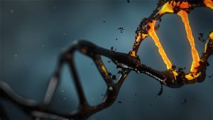
One of the ground breaking works dealt with the detection of UV induced changes in DNA based on the changes in water spectral pattern (Goto et al. 2015). This paper presents the discovery of non-invasive identification and measurement of very low concentrations of 3D conformations of DNA. It showed that even at small doses DNA structures water molecular system significantly different. The results of this paper suggested that, in contrast to DNA in solution (5–20 μ M), the formation of UV-induced cyclobutane pyrimidine dimers, CPD, cis-syn T<>Ts (0.77–3.0 μ M), caused increase of the strongly hydrogen bonded water, which has been found, in our previous studies, to be typical for oxidative stress. In addition, this paper showed that NIRS could qualitatively and quantitatively detect DNA and cis-syn T<>Ts in isolated DNA aqueous solutions upon UVC exposure even at these very low concentrations. For the first time, it has been shown that UV radiation (0-20kJ/m3) can be measured indirectly, by the changes induced in water spectral pattern, i.e. in the strength of the water covalent bonds and the conformation of water molecular system.
This paper opened a new venue for spectroscopic technologies and molecular biology and will provide revelatory insights into the biophysics and physico-chemical properties at the molecular level of life.
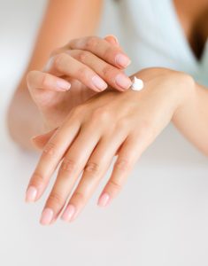
Aquaphotomics was also proposed for in vivo monitoring of topical cream effects (Matija et al. 2013, Matija et al. 2017), and the same holistic approach which was shown to be successful in water quality monitoring, was proposed for monitoring of dialysis efficacy (Munćan et al. 2016), which opens up new areas of aquaphotomics applications in the field of therapy monitoring.
Goto, N., G. Bazar, Z. Kovacs, M. Kunisada, H. Morita, S. Kizaki, H. Sugiyama, R. Tsenkova, and C. Nishigori. 2015. “Detection of UV-induced cyclobutane pyrimidine dimers by near-infrared spectroscopy and aquaphotomics.” Scientific reports 5.
Matija, L., J. Muncan, I. Mileusnic, and Dj. Koruga. 2017. “Fibonacci nanostructures for novel nanotherapeutical approach.” In Nano-and Microscale Drug Delivery Systems, 49-74. Elsevier.
Matija, L., R. Tsenkova, J. Munćan, M. Miyazaki, K. Banba, M. Tomić, and B. Jeftić. 2013. “Fullerene based nanomaterials for biomedical applications: engineering, functionalization and characterization.” Advanced Materials Research.
Munćan, J., I. Mileusnić, V. Matović, J. Šakota Rosić, and L. Matija. 2016. “The prospects of aquaphotomics in biomedical science and engineering.” Aquaphotomics: Understanding Water in Biology – 2nd International Symposium, Kobe University, Kobe, Japan, 26-29.12.2016.
Sakudo, A., R. Tsenkova, T. Onozuka, K. Morita, S. Li, J. Warachit, Y. Iwabu, G. Li, T. Onodera, and K. Ikuta. 2005. “A novel diagnostic method for human immunodeficiency virus Type‐1 in plasma by near‐infrared spectroscopy.” Microbiology and immunology 49 (7):695-701.